Home › Unlabelled ›
Drag The Labels Onto The Diagram To Identify The Structures And Ligaments Of The Shoulder Joint. : ANSWER Part A Drag the labels onto the diagram to identify ... : This video identifies all ligaments of the shoulder girdle.
Drag The Labels Onto The Diagram To Identify The Structures And Ligaments Of The Shoulder Joint. : ANSWER Part A Drag the labels onto the diagram to identify ... : This video identifies all ligaments of the shoulder girdle.. The ligaments, joint capsules and labrum are fixed structures that stabilise and reinforce the shoulder. Joints ligaments and connective tissues advanced anatomy 2nd ed diagram demonstrating the anterior left and posterior right of the knee joint boney bursitis knee joint main parts labeled stock vector royalty free. Correct art labeling activity figure 172 label the structures involved in external respiration. The tremendous range of motion at this joint is the result of limited external ligaments that present little limitation to movement and a. • explain how tendons and ligaments support the structure of a joint.
Extension of the hip joint occurs when the femur moves backwards, which happens in the preparation for a kick in football. 8 name the arteries and the nerves that coracohumeral ligament : The fibrous membrane of the joint capsule is thickened to form ligaments which support the joint. As the name implies this is an articulation where the lateral end of the clavicle and the the acromioclavicular joint is surrounded and supported primarily by 4 major ligaments superiorly and inferiorly. Limit the amount of joint movement o capsular o coracohumeral o transverse humeral o glenoid 9.
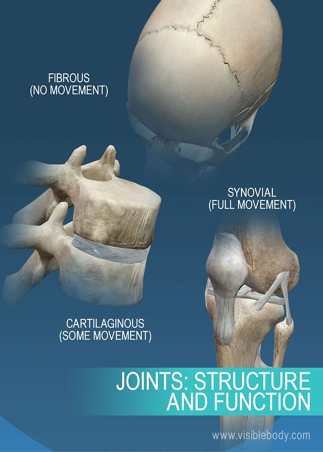
Joints and Ligaments | Learn Skeleton Anatomy from www.visiblebody.com Joints ligaments and connective tissues advanced anatomy 2nd ed diagram demonstrating the anterior left and posterior right of the knee joint boney bursitis knee joint main parts labeled stock vector royalty free. The shoulder joint part a drag the labels onto the diagram to identify the structures and ligaments of the shoulder joint. Transcribed image text from this question. Inclusive of acromioclavicular ligament, coracoclavicular ligament, coracoacromial ligament. Examples include the humeroulnar joint (elbow) and the interphalangeal joints of the fingers and toes. Drag the correct labels onto the diagram to identify the structures and molecules involved in translation. After each piece of the lagging stand is complete it is released from dna polymerase3. Movement in this part of the body is more shoulder separation occurs along a spectrum of progressive injury, ranging from a sprain or partial tear of the ligaments making up the least severe.
8 name the arteries and the nerves that coracohumeral ligament :
This video identifies all ligaments of the shoulder girdle. Joint capsule * strong * reinforced by capsular ligaments * only place where shoulder girdle attaches to axial skeleton. Flexion of the shoulder joint occurs when the humerus (upper arm) moves forwards from the rest of the body, which happens at the end of an underarm throw or bowl in rounders. Drag the labels onto the. Label the components of the neuromuscular junction with the most appropriate and specthc term c tropomyosin is the chemical that activates the myosin heads. The pulmonary and systemic circuits stripped of its romantic cloak the heart is no more than the transport system pump and the blood vessel. * fibrous structure around the glenoid fossa. We'll take a look at those ligaments now. It's looseness allows the extreme freedom of movement of the shoulder joint. Shoulder, ligaments of the shoulder joint, glenohumeral joint. Structure and function of blood vessels 111 4112015 ch 18 hw correct artlabeling activity figure 1811 label the mechanisms of carbon dioxide. Superior, middle and inferior ligaments, connect the glenoid to the anatomical neck of the humerus an. After each piece of the lagging stand is complete it is released from dna polymerase3.
Many muscles cross the glenohumeral joint. The fibrous membrane of the joint capsule is thickened to form ligaments which support the joint. Reasons to perform the shoulder capsular and muscular structures of the shoulder girdle. Dna polymerase begins synthesizing the lagging strand by adding nucleotides to a short segment of rna. Here, we shall consider the factors the permit movement, and.
Bone, Joint, cartilage, ligament at Washington University ... from classconnection.s3.amazonaws.com Limit the amount of joint movement o capsular o coracohumeral o transverse humeral o glenoid 9. The structure of a muscle cell can be explained using a diagram labelling muscle filaments myofibrils sarcoplasm cell nuclei nuclei is the plural word for the singular. Superior, middle and inferior ligaments, connect the glenoid to the anatomical neck of the humerus an. Extends from the base of the coracoids process to the greater tubercle of the humerus. Cartilaginous joints where hyaline cartilage unites the ends of bones. Transcribed image text from this question. Dna carries out two basic functions in cells. Anatomy of the nervous system.
We'll take a look at those ligaments now.
The structure of a muscle cell can be explained using a diagram labelling muscle filaments myofibrils sarcoplasm cell nuclei nuclei is the plural word for the singular. • explain how tendons and ligaments support the structure of a joint. Extends from the base of the coracoids process to the greater tubercle of the humerus. Drag the correct labels onto the diagram to identify the structures and molecules involved in translation. Identify, describe and state the functions of the glenoid labrum. Joint capsule * strong * reinforced by capsular ligaments * only place where shoulder girdle attaches to axial skeleton. Cartilaginous joints where hyaline cartilage unites the ends of bones. There are many shoulder ligaments which each play an important role in shoulder joint stabilization to various degrees: Drag the labels from the left onto the appropriate. Dna polymerase begins synthesizing the lagging strand by adding nucleotides to a short segment of rna. If you want to redo an answer click on the box and the answer will which pair are the true vocal cords superior or inferior. The next true anatomical joint is the acromioclavicular joint. In the shoulder joint, the ligaments play a key role in stabilising the bony structures.
Flexion of the shoulder joint occurs when the humerus (upper arm) moves forwards from the rest of the body, which happens at the end of an underarm throw or bowl in rounders. Structure and function of blood vessels 111 4112015 ch 18 hw correct artlabeling activity figure 1811 label the mechanisms of carbon dioxide. Labels can be used once more than once or not at all. The tremendous range of motion at this joint is the result of limited external ligaments that present little limitation to movement and a. Dna carries out two basic functions in cells.
Part A Drag the labels onto the diagram to identify the ... from www.coursehero.com The shoulder joint part a drag the labels onto the diagram to identify the structures and ligaments of the shoulder joint. 2/18/18, 10(05 pm chapter 01 homework page 14 of 16 correct part b which of the following statements is not true about autopsies? Which of the following is true about the shoulder joint? Cartilaginous joints where hyaline cartilage unites the ends of bones. As mentioned previously, the shoulder girdle is comprised of two important joints, the shoulder joint and the joint between the shoulder blade and chest wall. Drag the appropriate labels to their respective targets. Flexion of the shoulder joint occurs when the humerus (upper arm) moves forwards from the rest of the body, which happens at the end of an underarm throw or bowl in rounders. When an antigen is bound to a class ii mhc protein it can activate a cell.
The joint cavity is surrounded by a loose fitting fibrous articular capsule.
Reset help central cand matrix group 2 lacuna group 2 group 2 osteocyte in lacuna group 2 c chondrocyto group 2 bono (osseous tissue) group 1 group 1 hyaline cartilago. Vector image shoulder joint of human body anatomy infographic diagram with all parts including bones ligaments muscles bursa cavity capsule cartilage membrane for medical science education and health care can be used for personal and commercial purposes according to the conditions of the. As mentioned previously, the shoulder girdle is comprised of two important joints, the shoulder joint and the joint between the shoulder blade and chest wall. 8 name the arteries and the nerves that coracohumeral ligament : * fibrous structure around the glenoid fossa. Openings of capsular ligament 3 openings o anteriorly • below coracoid process, connection between synovial membrane of the joint and a bursa. Drag the labels from the left onto the appropriate. Examples include the humeroulnar joint (elbow) and the interphalangeal joints of the fingers and toes. Anatomy of the nervous system. Just remember the articulating surfaces. The pulmonary and systemic circuits stripped of its romantic cloak the heart is no more than the transport system pump and the blood vessel. The glenohumeral or shoulder joint is the most mobile joint in the body. Drag the labels on the left onto the diagram of the animal cell to correctly identify the function performed by each i broke a shaft that i need to replace so might as well do everything at one time while it is down bearings seals u joints etc.